Anne-Dominique Gindrat is a postdoc in the Neurobiology Laboratory of Prof. Hansjörg Scherberger at the Deutsches Primatenzentrum GmbH in Göttingen. She is investigating some brain areas involved in planning and executing hand grasping movements in behaving macaque monkeys using the emerging approach of optogenetics.
PhD in Natural Science, option Neuroscience, 2015
University of Fribourg (Switzerland)
MSc in Biology, 2010
University of Fribourg (Switzerland)
BSc in Biology, 2008
University of Fribourg (Switzerland)
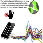
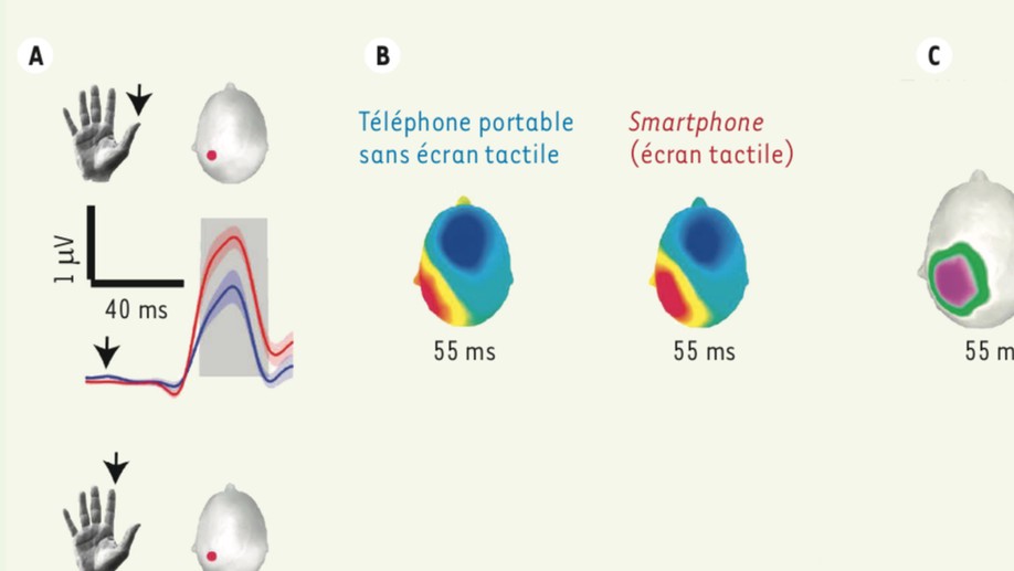
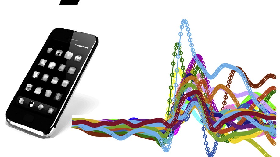
Cortical activity allotted to the tactile receptors on fingertips conforms to skilful use of the hand. For instance, in string instrument players, the somatosensory cortical activity in response to touch on the little fingertip is larger than that in control subjects. Such plasticity of the fingertip sensory representation is not limited to extraordinary skills and occurs in monkeys trained to repetitively grasp and release a handle as well. Touchscreen phones also require repetitive finger movements, but whether and how the cortex conforms to this is unknown. By using electroencephalography (EEG), we measured the cortical potentials in response to mechanical touch on the thumb, index, and middle fingertips of touchscreen phone users and nonusers (owning only old-technology mobile phones). Although the thumb interacted predominantly with the screen, the potentials associated with the three fingertips were enhanced in touchscreen users compared to nonusers. Within the touchscreen users, the cortical potentials from the thumb and index fingertips were directly proportional to the intensity of use quantified with built-in battery logs. Remarkably, the thumb tip was sensitive to the day-to-day fluctuations in phone use.
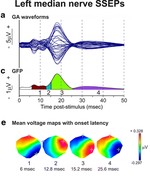
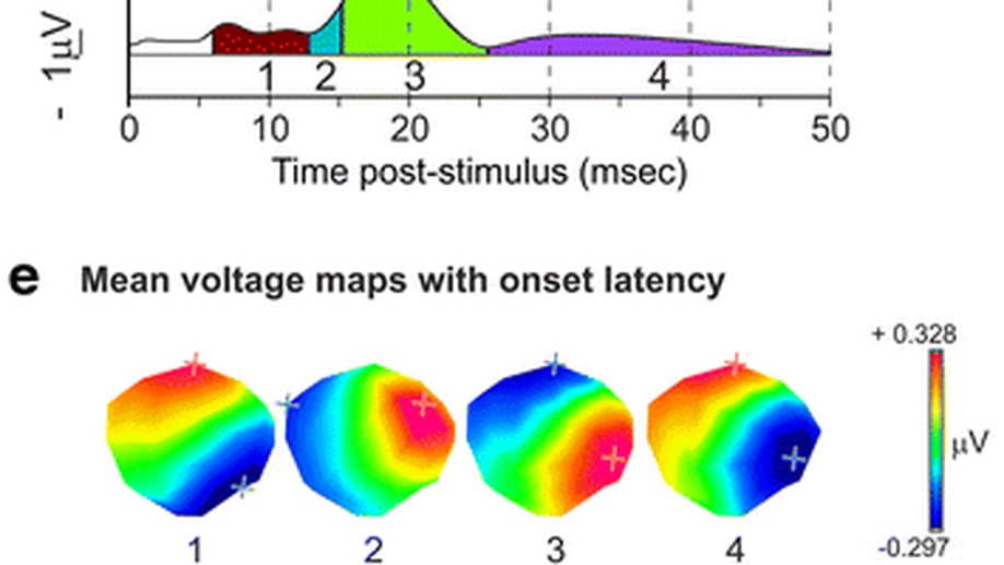
High-density scalp EEG recordings are widely used to study whole-brain neuronal networks in humans non-invasively. Here, we validate EEG mapping of somatosensory evoked potentials (SSEPs) in macaque monkeys (Macaca fascicularis) for the long-term investigation of large-scale neuronal networks and their reorganisation after lesions requiring a craniotomy. SSEPs were acquired from 33 scalp electrodes in five adult anaesthetized animals after electrical median or tibial nerve stimulation. SSEP scalp potential maps were identified by cluster analysis and identified in individual recordings. A distributed, linear inverse solution was used to estimate the intracortical sources of the scalp potentials. SSEPs were characterised by a sequence of components with unique scalp topographies. Source analysis confirmed that median nerve SSEP component maps were in accordance with the somatotopic organisation of the sensorimotor cortex. Most importantly, SSEP recordings were stable both intra- and interindividually. We aim to apply this method to the study of recovery and reorganisation of large-scale neuronal networks following a focal cortical lesion requiring a craniotomy. As a prerequisite, the present study demonstrated that a 300-mm(2) unilateral craniotomy over the sensorimotor cortex necessary to induce a cortical lesion, followed by bone flap repositioning, suture and gap plugging with calcium phosphate cement, did not induce major distortions of the SSEPs. In conclusion, SSEPs can be successfully and reproducibly recorded from high-density EEG caps in macaque monkeys before and after a craniotomy, opening new possibilities for the long-term follow-up of the cortical reorganisation of large-scale networks in macaque monkeys after a cortical lesion.